
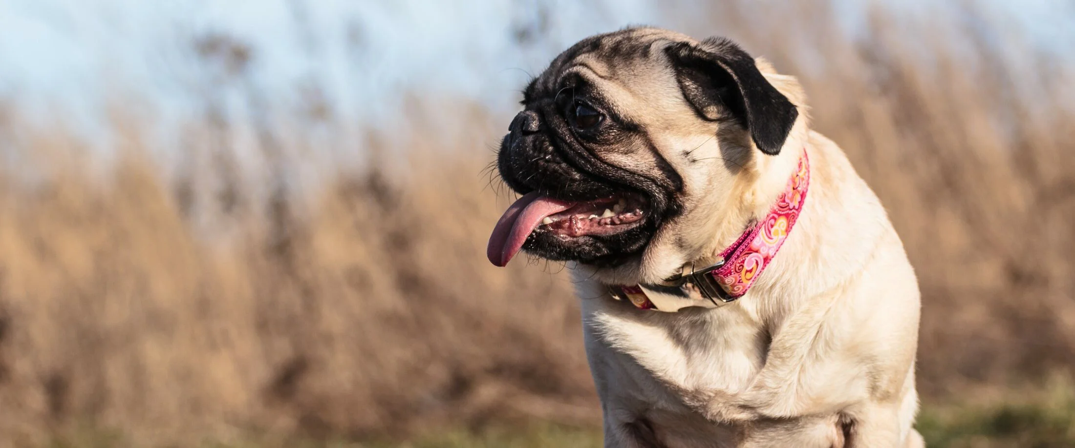
In-House Diagnostic Tests
This entails taking samples from the ear canal (usually with a cotton swab), rolling the samples on a microscope slide, then heat fixing and staining the samples, and then evaluating the sample under a microscope. The samples can often tell us what type of infection is present in the ear, how to treat infection more effectively and if the infection is resolving or has resolved.
Ear Swabs for Mites
Swabs are typically taken from the ears and rolled onto a microscope slide with mineral oil and evaluated under a microscope.
Skin Cytologies
This entails taking samples from the skin’s outer layers, staining them with a special stain, and evaluating them under a microscope for infection, inflammation, tumors, and so on.
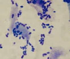
Skin cytology sample
Skin Scrapings
A procedure that involves gently scraping the surface of the skin with a dull blade or special medical spatula and mineral oil to collect the sample(s). The sample is then examined under a microscope for mites that live on the skin or in hair follicles. Hair from an area that needs scraping may need to be shaved at times.
Trichograms
Trichograms are evaluating the hairs under the microscope to look for certain infections, trauma and/or malformation. Trichograms can sometimes also be used to evaluate for mites that live in hair follicles (Demodex mites).
Wood’s Lamp Examination
Wood’s lamp examination uses ultraviolet light to evaluate for fluorescence seen with some ringworm infections. FYI ringworm is a fungus, not a worm.